Interview with Louise Breideband: Brighter’s PhD student at GUF

Brighter is a European Project that brings together different academic and industrial partners to develop a new 3D bioprinting technology able to produce human tissues at high speed and with high spatial resolution. This innovative technology is based on light-sheet lithography and an original top-down approach.

Louise Breideband is a PhD student in the frame of Brighter Project at the Physical Biology laboratory in the Buchmann Institute for Molecular Life Sciences, Goethe University Frankfurt.  After a master’s degree in food production and a couple of years doing scientific communication, she decided to come back to the lab! She completed a master’s degree in physical biology of cells and cell interactions, and now she is performing a PhD and contributing to Brighter project in the development of a 3D bioprinter using light sheet photopolymerization. Get to know more about Louise in this interview!

Can you describe yourself in a couple of lines?

My name is Louise Breideband, I am a PhD student in the Physical Biology lab in the BMLS, Goethe University Frankfurt. Our lab is specialized in microscopy, especially light-sheet microscopy and high-resolution imaging. I am originally French and graduated with a master’s degree in food production from the university of AgroParisTech (Paris, France). I worked for a couple of years in Australia and Germany as a scientific communication expert, but I soon realized that I was missing hands-on work in a laboratory. I completed a master’s degree in physical biology of cells and cell interactions in the Goethe University Frankfurt before joining Professor Stelzer’s lab for my master thesis and PhD thesis. Under Dr. Pampaloni’s supervision, I am working closely with Levin Hafa on developing a 3D bioprinter using light sheet photopolymerization. Additionally, I am assisting on a project developing a 3D model of bone marrow using spheroids that will be exposed to microgravity and sent to the international space station in 2022.

What is your role/position within Brighter?

I am at the interface between the different participants in the project, testing various hydrogel compositions delivered by Cellendes and encapsulating fibroblasts following protocols developed by IBEC and Technion, printed on the 3D bioprinter prototype built by us in cooperation with Mycronic.

Could you tell us a little bit about the concrete work you’re involved in inside Brighter project?

I am confirming that results established by IBEC, Technion or Cellendes still apply when using our innovative 3D bioprinter. For example, my first task was to determine how the hydrogel was polymerizing when initiated with a light-sheet, which I demonstrated using fluorescent dye diffusion and FRAP (fluorescent recovery after photobleaching). Then, I tested the cell viability of fibroblasts printed in various hydrogel compositions and imaged the cells in live under a light sheet microscope for 7 to 9 days. Finally, using the patterning developed by Levin Hafa and Mycronic, we printed the cells in a more intricate design, showcasing the full capacity of the prototype. The next step would be to co-culture the fibroblasts and the keratinocytes in the printed constructs.
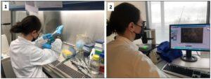
1- Mixing the hydrogel and the cells before printing.

2- Looking at cells under the microscope. Fibroblasts were dyed with a live fluorescent dye so that we can observe the cells after 3D bioprinting.


What are the expected results?

We expect the cells to grow, multiply and express markers when 3D bioprinted as fibroblasts would do in vitro. Early results are very encouraging in this regard. Eventually, we would like to develop a functional 3D bioprinter that is suitable to encapsulate cells in high definition while mimicking the in vitro environment.

What is the expected impact of the work you’re doing?

At Goethe University Frankfurt, we are at the crossroad of all our colleagues in the partner institutions. We closely work with them to make sure that all the intricate parts of this project come together. Currently, we have the only prototype in the project which makes us responsible for delivering results using this prototype, as well as further developing it and sharing our observations with our partners, building a feedback loop that fully exploits this collaboration.

How do you feel about being a part of this European Project?

As a product of Europe myself, I feel very proud to be involved in this journey. I love working on a multi-disciplinary project which allowed me to professionally grow in the last two years. Unfortunately, because of the pandemic, it has been very difficult to physically interact and visit each other, which is a real shame. However, online communication flourished and thanks to video calls, we are still able to see each other and reach out on a regular basis.

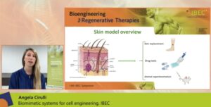
 

Angela Cirulli shows BRIGHTER advances at the 14th IBEC Symposium

Last 27th and 28th of October took place the 14th IBEC Symposium, completely online due to COVID-19 pandemic restrictions. This year, the event focused on Regenerative Therapies and how the latest scientific advances in mini-organs, organs on a chip, 3D bioprinting and tissue engineering can help to address the problem of tissue demand for organ transplantation and replacement, and for experimentation for new drugs development. The symposium counted with the participation of more than 300 registered attendees, including renowned speakers from Europe and the USA as Jennifer A. Lewis from Harvard University, Frank P. Luyten, director of the Skeletal Biology and Engineering Research Center at Belgium, and Eva Van Rooij from the Hubrecth Institute in The Netherlands.

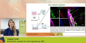
In the frame of this symposium, Angela Cirulli, predoctoral researcher at the Biomimetic Systems for Cell Engineering from the Institute for Bioengineering of Catalonia (IBEC), presented the last advances of the BRIGHTER project on 3D bioprinting and the development of a skin model. She presented a poster on a virtual space and was one of the 17 researchers selected to show her work also on a flash talk. Concretely, she talked about “Photo-crosslinkable hydrogels for skin model reconstruction” and presented the latest results regarding hydrogels obtained at the Biomimetic Systems for Cell Engineering group at IBEC in close collaboration with one of the industrial partners of the project, Cellendes.

Angela presented BRIGHTER’s novel approach based on visible-light 3D bioprinting system to produce skin tissue models as similar as possible to a real structure. One of the main limitations of working with skin tissue engineered models is to obtain a faithful replica of this heterologous and complex organ that could be used for skin replacement and drug testing (avoiding animal experimentation) among other purposes. In this context, photo-crosslinkable hydrogels are the best candidates to mimic the perfect skin cells microenvironment, due to their biocompatibility and their tuneable properties. In BRIGHTER project, researchers are using norbornene based hydrogels with the aim to construct a skin model with improved properties.

She explained the polymerization process and presented immunostaining results showing the formation of two different cellular compartments, mimicking both the dermis (top layer) and the epidermis (bottom layer). To do so, representative skin cellular types were included on the hydrogels (some cells seeded on top and others included within) and co-cultured for several days, showing a high cell viability and uniform distribution. Moreover, she also described a higher production of extracellular matrix proteins of co-cultured inside the hydrogel, revealing crosstalk among them.


“These promising results do not only demonstrate the good compatibility between cells and the hydrogel structure but are also tracing an excellent pattern to reach a final skin model”

Angela Cirulli

Interview with Anna Altshuler: postdoctoral researcher at Technion

BRIGHTER is a European Project that brings together different academic and industrial partners to develop a new 3D bioprinting technology able to produce human tissues at high speed and with high spatial resolution. This innovative technology is based on light-sheet lithography and an original top-down approach.

Anna Altshuler is a Postdoctoral researcher working in the frame of BRIGTHER Project at Technion, Israel.  She has a bachelor’s degree in biotechnology engineering and has a strong background in the study of stem cells using different approaches and techniques. Her previous research experience adds, undoubtedly, high value to the project! Discover in the interview below some aspects of her personal and professional profile, as well as her role in BRIGHTER project.


Can you describe yourself in a couple of lines?

My name is Anna Altshuler, a Postdoctoral researcher in Ruby Shalom-Feuerstein lab at Technion. Our lab is interested in epithelial stem cells and pathophysiology focusing on skin and cornea. I was born in Ukraine and my family immigrated to Israel when I was a child. After graduating from high-school I completed my military service in the IDF (Israel Defense Forces) as a computer network manager. Following my army service, I did a bachelor’s degree in biotechnology engineering at Braude college. For my master and PhD degrees I joined Ruby Shalom-Feuersteins’ lab at Technion. In my master studies, I focused on the role of RAS oncogene on pluripotent stem cells. I worked mainly with mouse pluripotent stem cells and many molecular techniques like RT-PCR, western blot and immunofluorescent staining. In my PhD I focused more on the adult stem cells of the cornea by using different genetic mouse models and advanced genomic technologies as single cell RNA sequencing.


What is your role within Brighter?

My role in the BRIGHTER Project is trying to use the knowledge I gained on stem cells by growing them on the gels producing the differentiated cells that are a part of functional 3D skin tissue.

Could you tell us a little bit about the concrete work you’re involved in inside Brighter project?

I am currently working on integrating primary human cells isolated in our lab with the new gel technology in order to test their ability to survive, migrate and proliferate inside the gel. By using known cellular markers, we will be able to see follow the evolution of the different cell types forming a skin model.

What is the expected impact of the work you’re doing?

Our results should demonstrate the proof-of-concept for BRIGHTER bioprinting technology combined with different kinds of primary cells that form the skin tissue.

How do you feel about being a part of this European Project?

Since the pandemic, I didn’t have the chance to meet my fellow project partners in person. It’s always a pleasure to meet them on “Zoom” and discuss the interesting results or consult about different issues in the project.

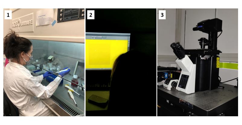
Overview of an experiment to prepare light inducible hydrogels containing cells.
1- Gels are mixed with mouse fibroblasts cells that are known to support primary keratinocytes
2- I adjust the settings for gel induction according to our experimental design
3- Finally, gels are illuminated under this special microscope to polymerize


Interview with Gustaf Mårtensson: Brighter’s project leader at Mycronic

Brighter is a European Project that brings together different academic and industrial partners to develop a new 3D bioprinting technology able to produce human tissues at high speed and with high spatial resolution. This innovative technology is based on light-sheet lithography and an original top-down approach.

Gustaf Mårtensson has a background in fluid mechanics and works at Mycronic, a Swedish high-tech company that has been active in the electronics industry for more than 30 years. He works connecting technological solutions from Mycronic with new applications, and he is also adjunct researcher at Royal Institute of Technology (KTH) in Stockholm, Sweden, within clinical diagnostics. In summary, one of his main tasks is to connect people among them. You can check in this video what his daily life at work looks like! You can also read the interview to get to know more about Gustaf and his participation in BRIGHTER project. Enjoy it!

Can you describe yourself in a couple of lines?

I’m Gustaf Mårtensson and I have a background in fluid mechanics from Royal Institute of Technology in Stockholm, Sweden. I work in technology development finding new technological solutions for our applications, but also new applications for our technologies. I’m also adjunct researcher at Royal Institute of Technology (KTH) in Stockholm, Sweden, within clinical diagnostics.

What is your role/position within Brighter?

I’m Project leader together with Robert Eklund for the BRIGHTER project at Mycronic and I organise the work of all our experts in optics, lasers and data handling.

Could you tell us a little bit about the concrete work you’re involved in inside Brighter project?

Our work is focused on the actual patterning technology together with Göethe Universität in Frankfurt with Francesco, Louise, Sven and Levin. The technology will be able to address individual spatial elements (voxels) in the hydrogel which means that we have to be able to control the laser light in space in time. Mycronic’s part of the device is scanning the laser in one direction and controlling the intensity in time using acousto-optical methods. This will be combined with GUFs module that will scan in an orthogonal direction and house the sample in an appropriate environment.

What are the expected results?

The outcome of our work is the combination of the two modules resulting in the actual patterning device, which together with the work of Cellendes, IBEC and Technion will result in digitally defined patterned tissue.

How do you feel about being a part of this European Project?

This project is one of the highlights of my work week. Sure, it’s challenging, but that’s what makes it fun. I get to work with super talented people at Mycronic and I get to collaborate with great people at GUF, Cellendes, IBEC, and Technion!


Interview with Gabriele Di Napoli: Chemical Technical Assistant at Cellendes

Brighter is a European Project that brings together different academic and industrial partners to develop a new 3D bioprinting technology able to produce human tissues at high speed and with high spatial resolution. This innovative technology is based on light-sheet lithography and an original top-down approach.

Gabriele Di Napoli is a Chemical Technical Assistant in the frame of Brighter Project working at Cellendes. He has a degree as a Chemical Engineer and for the last years he has been applying his knowledge in biomedicine, in the study of tumours or Parkinson’s disease. Now he joined Brighter Project to the develop and prepare photo-crosslinkable hydrogels and study their compatibility with cell culture. You can check here a short video where he briefly shows one of the experiments he usually performs in the lab., and an interview where he explains in more detail some aspects of his professional profile and his role in Brighter project. Enjoy it!

Can you describe yourself in a couple of lines?

Hi, my name is Gabriele Di Napoli, I come, like the name says, from Naples, Italy. I have a degree as a Chemical Engineer. I have been studying Chemistry at the university for two years and after that I started to work at the National Council of Research (CNR) in Naples. In the first year I have been involved in the study of the angiogenesis in tumor development and then I moved to another group studying mouse embryonic stem cells. After 6 years I moved to Tübingen for working for 2 years in a group involved in the study of Parkinson´s disease using iPSCs. Now it has been almost 2 years that I am working at Cellendes as Chemical Technical Assistant.

What is your role/position within Brighter?

I am involved in the study, development and preparation of photo-crosslinkable hydrogels, including their compatibility with cell culture.

Could you tell us a little bit about the concrete work you’re involved in inside Brighter project?

I do the chemical modification of polymers, and the evaluation of the chemical/physical properties using methods like HPLC, spectrophotometry or rheology.

What are the expected results?

We are working towards getting cell-compatible hydrogels that can be formed by light illumination and that can be modified according to the need of different cell types. Another aspect of our work is to make sure that the cells don’t sink to the bottom during the set up of the hydrogels. We also would like to be sure that the set up of the hydrogels is simple and robust enough to enable our project partners to use them safely and reliably.

What is the expected impact of the work you’re doing?

In my opinion this project will provide many interesting results which could be very useful for the scientific community. It is important to be able to fabricate spatially and chemically highly defined bioprinted hydrogels to have better tools to study tissue organization and development. It is almost equally important to communicate the results properly to make sure that this development will be continued in the future.

How do you feel about being a part of this European Project?

Working with different groups around Europe is exciting. For me it is important to have contact with the other partners, to collaborate with them on different experiments and to compare or confirm the results. Furthermore, it is very interesting to get to know people from different corners of Europe and to see how they work.


Discovered two limbal stem cell populations that control corneal homeostasis and wound healing processes

Ruby Shalom-Feuerstein, Anna Altshuler and Aya Amitai-Lange, the workforce of Brighter project at Technion, Israel, led a work that has just been published in the prestigious journal Cell Stem Cell. They describe for the first time the existence of two limbal stem cell populations that have different characteristics and functions in the maintenance of the cornea. This pioneering study opens the doors to the development of accurate treatments for corneal disorders.

Limbal epithelial stem cells (LSCs) are responsible for the renewal of the cornea and up to date, have been seen by the scientific community as rare and slow-cycling cells. Now, a team or researchers from different laboratories at Technion – Israel Institute of Technology have discovered that in fact, the LSCs are composed by two different populations of cells, with distinct characteristics and niches: the quiescent and the active limbal stem cells. The work, led by Ruby Shalom-Feuerstein and signed by Anna Altshuler and Aya Amitai-Lange as first authors, has been recently published in the journal Cell Stem Cell. As experts in the epithelial stem cells field, these researchers from the Laboratory of Epithelial Stem Cells & Pathophysiology at Technion, are in charge to provide the cellular models and scientific knowledge about stem cell biology to BRIGHTER Project and to collaborate on the fabrication and testing of the new skin engineered tissues using the biomaterial scaffolds developed by the bioengineering partners.


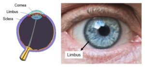
The limbus is a ring-shaped zone at the corneal-conjunctival boundary that hosts the corneal epithelial stem cells, that is, the cells responsible for the renewal of old corneal cells and of wound healing response. Thus, a region extremely important for the proper functioning of the eyes.


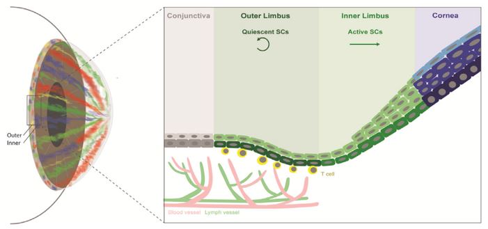
Combining single cell RNA sequencing and quantitative lineage tracing techniques in mouse, researchers discovered the existence of two populations of abundant limbal epithelial stem cells (LSCs) localized in separate and well-defined sub-compartments, termed the ‘‘outer’’ and ‘‘inner’’ limbus. The quiescent LSCs (qLSCs) are found in the outer limbus zone, co-localize with and regulated by T-cells. qLSCs participate in wound healing and boundary formation. On the other hand, the active LSCs (aLSCs) that reside in the inner limbus are responsible for the cornea homeostasis and renewal.

The observation that there are two populations of stem cells, and that these cells are abundant, challenges the convectional LSC dogma, that considered a single rare LSC population. Nevertheless, this conclusion fits with the equipotent stem cell model, describing epithelial stem cell dynamics in the gut, epidermis and the oesophagus.

The complete atlas of the murine corneal epithelial lineage presented in this work represents a highly valuable tool to understand eyes disorders and help finding preventive and curative treatments for patients that suffer from corneal blindness due to LSC deficiency. In summary, the new data provided in this study pave the way to the development accurate bioengineering tools, as high-quality organoids, better LSC-based therapies, and application of LSCs in regenerative medicine.


Reference article: Discrete limbal epithelial stem cell populations mediate corneal homeostasis and wound healing. Anna Altshuler; Aya Amitai-Lange; Noam Tarazi; Sunanda Dey; Lior Strinkovsky; Shira Hadad-Porat; Swarnabh Bhattacharya; Waseem Nasser; Jusuf Imeri; Gil Ben-David; Ghada Abboud-Jarrous; Beatrice Tiosano; Eran Berkowitz; Nathan Karin; Yonatan Savir and Ruby Shalom-Feuerstein. Cell Stem Cell 28, 1–14, 2021.


Interview with Levin Hafa: Brighter’s PhD student at GUF

Brighter is a European Project that brings together different academic and industrial partners to develop a new 3D bioprinting technology able to produce human tissues at high speed and with high spatial resolution. This innovative technology is based on light-sheet lithography and an original top-down approach.

Levin Hafa is a PhD student working at the Buchmann Institute for Molecular Life Sciences  (BMLS) at Goethe University Frankfurt (GUF) in the frame of Brighter. His is a biologist specialized in molecular and cell biology with experience in organoid and organ-on-chip technology, stem cells, human liver models and microfluidic devices. In this short video we can see one his tasks in the project related with the hydrogels patterning process. In this interview Levin explains his professional profile, formation, and his role in Brighter project. For sure his knowhow will be very important to the success of the project!

Can you describe yourself in a couple of lines?

I am Levin Hafa, a PhD student at the physical biology group of Prof. Ernst Stelzer at the Goethe University in Frankfurt, Germany. I am from Germany and originally from close to Frankfurt as well. After graduating from high school, I studied Biology at the Technical University of Darmstadt (TU) for my bachelor’s with a focus on molecular and cell biology. I then decided to pursue my master’s degree at the TU Darmstadt and joined the M.Sc. Technical Biology program. During my Erasmus semester in Barcelona at Universitat de Barcelona, I was first introduced into Organoid and Organ-on-chip technology. For my master’s thesis, I moved to Berlin and worked with stem cells, human liver models and microfluidic devices at TissUse, a TU Berlin spin-off. Being inspired by Tissue Engineering, Organ-on-chip technology and the 3R principles I was looking for a challenging PhD position in this field of biology. At Goethe University Frankfurt and with the BRIGHTER project in particular, I found the perfect environment to play to my strengths and pursue my interests.

What is your role/position within Brighter?

One of my main tasks within the BRIGHTER project is to develop and validate a patterning mechanism for 3D-bioprinting in the light-sheet (LS) system. Once this task is achieved, I will use this knowledge to complete my PhD project developing a 3D printed human liver model to establish another proof-of-concept for our bioprinting platform.

Could you tell us a little bit about the concrete work you’re involved in inside Brighter project?

For the purpose of creating a patterning mechanism, I need to connect the fields of polymer chemistry, optics, software engineering and cell biology. Specifically, I am designing code, which interprets G-Code, a common 3D Printer language, which is able to transform a 3D CAD rendered model into lines of code and back to a 3D print. I then validate the written code by printing complex three dimensional structures with photosensitive hydrogels. Currently, I am also training to validate the code by printing 3D structures with embedded cells.

What are the expected results?

Once the patterning is established, we expect it to be able to recreate complex 3D structures both protruded and hollow features, focusing special attention on developing thin channels, as observed in human vasculature. Furthermore, we could recreate stem-cell niches, to help a model organ sustenance for a long period of time.

What is the expected impact of the work you’re doing?

My work is especially important for BRIGHTER’s project, since it is the connection between our current LS system and the patterning system developed by our partner Mycronic, allowing for the 3D bioprinting of complex structures, that resemble the human physiology. Additionally, a quick way to 3D-print functional and long-lasting human tissue models would be of great interest for many pharmaceutical companies in terms of toxicological studies and preclinical trials.

How do you feel about being a part of this European Project?

Since I started my PhD in mid-2020, due to the pandemic I was not able to meet my fellow project partners in person yet. But I really enjoy working within an international, interdisciplinary team of scientists and can not wait to visit them.


BRIGHTER Project awarded with the best video-poster prize at BioNTERM Conference

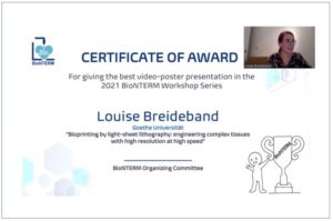
BRIGHTER European Project attracted attention and stood out at the last BioNTERM conference: Bio and Nanomaterials in Tissue Engineering and Regenerative Medicine. Louise Breideband, PhD student at the Buchmann Institute for Molecular Life Sciences at GUF (Goethe University Frankfurt), presented the project on a video-poster where she explained the objectives and the new 3D bioprinting technique being developed. This video was awarded with the best video-poster prize, recognizing the quality of the presentation and the scientific content. In addition, confirming the interest aroused, she was also selected to give a flash talk about BRIGHTER during the event.

BioNTERM conference was the first Virtual Conference and Workshop Series on Bio and Nanomaterials in Tissue Engineering and Regenerative Medicine taking place in Canada. The event, at the interface of Bio and Nanomaterials, brought together experts in different fields from Scientific and Science Communication to Translational Medicine and Regenerative Therapies to boost the integration of different disciplines ranging from materials engineering to clinical practices in order to accelerate discovery and clinical translation.

The BioNTERM conference was held completely online during the five March Monday afternoons of 2021 (1st, 8th, 15th, 22nd and 29th). This innovative format aimed to increase the interaction among participants and speakers, and to maximize learning.


More information: Home | BioNTERM


Louise Breideband presents BRIGHTER at the first Future 3D Additive Manufacturing, the 3DMM2O Conference 2021

During the first Future 3D Additive Manufacturing – the 3DMM2O Conference, held online last 1st to 4th of March, Louise Breideband, PhD student at the Buchmann Institute for Molecular Life Sciences at GUF (Goethe University Frankfurt), presented the BRIGHTER project on a flash talk and a poster, both signed by all partners of the project.

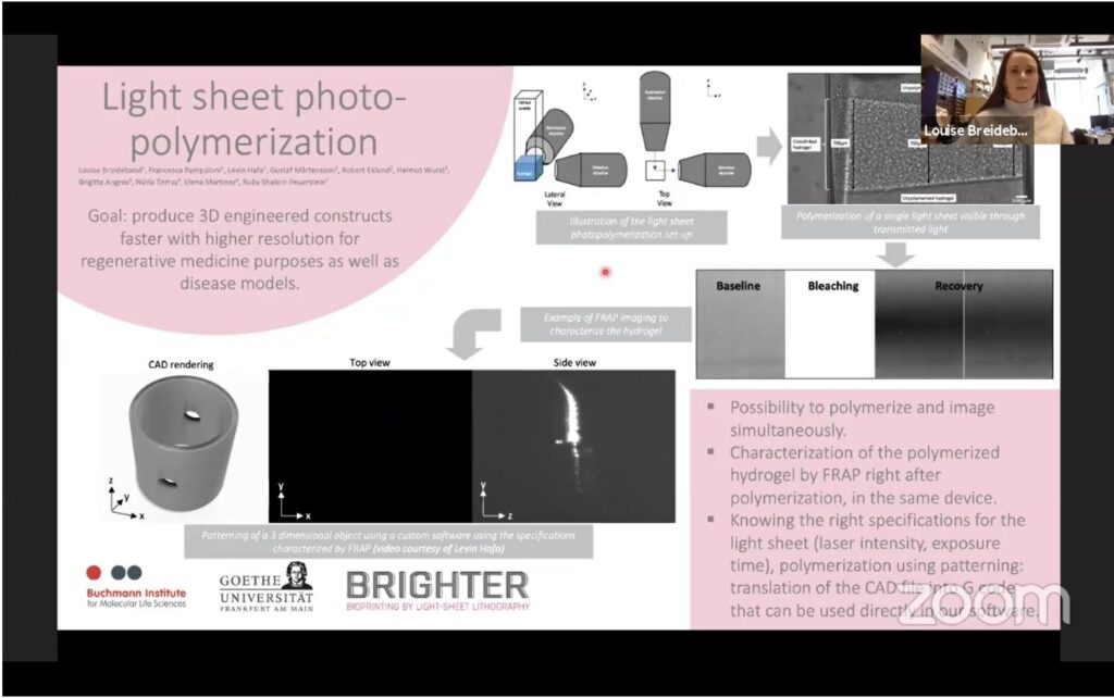
Louise explained the main objectives of BRIGHTER in the talk, entitled “Light sheet photopolymerization”, that are to increase 3D bioprinting resolution and speed for biomedical applications, and also mentioned the future steps towards the development of this new technology.

Additionally, she presented a poster showing more details related with this new technology and summarizing the last achievements of the project.


3DMM2O (“3D Matter Made to Order”) is a Cluster of Excellence, a joint Research Cluster of Karlsruhe Institute of Technology (KIT) and Heidelberg University (Uni HD), that organizes an annual conference on topics surrounding 3D Additive Manufacturing. The aim of the conference is to provide the latest research developments and future trends in 3D nano- and micro-manufacturing technologies and its applications in industry and research, to break barriers of scale, precision and speed.

More information: Future 3D Additive Manufacturing – the 3DMM2O Conference


Members of Brighter project at the Physical Biology Group talk about light-sheet microscopy and bioprinting

Francesco Pampaloni, Louise Breideband and Levin Hafa recently participated in a video to explain the work developed at the Physical Biology Group from the Buchmann Institute for Molecular Life Sciences at the Goethe University Frankfurt (GUF). Their role in Brighter Project is, together with Mycronic, to adapt the light-sheet microscopy technique to 3D bioprinting. In their laboratory they are focused in applying physical models and physical technologies to understand biological systems.

Brighter project is developing a new 3D bioprinting technology based in light-sheet microscopy, and the expected outcome is to have a technology capable of producing engineered human tissues with a high spatial resolution and at a high printing speed. Moreover, the use of the light-sheet microscopy will allow to observe in real time the fabrication of the tissue while it is being printed.

To achieve this ambitious aim, five academic and industrial partners are involved in Brighter project, and people from Goethe University Frankfurt (GUF) have a crucial role in the constructions of the 3D bioprinter equipment. The Physical Biology Group, led by Prof. Dr. Ernst H.K. Stelzer, is a pioneer in the field of Light Sheet Fluorescence Microscopy applied to cell and developmental biology. They employ physiologically relevant cellular models for both fundamental and applied as well as translational research.

Dr. Francesco Pampaloni, Louise Breideband and Hevin Hafa are the researchers from this lab involved in Brighter project. Recently they participated in a video created in the context of the Giersch – Summerschool & International Conference 2021 where they talk about light-sheet microscopy and bioprinting (from 1’48”).

At the Physical Biology Group they apply physical methods, in particular light and fluorescence microscopy to analyze biological specimens in 3D, concretely, in the frame of Brighter project the main tool they are developing is the light -sheet fluorescence microscopy (LSFM).

According to Francesco Pampaloni, the main advantages of this technique is that with a minimal exposure of the samples to light, so they are preserved for a longer time, it is possible to have very fast and precise 3D imaging of a specimen.

On the other hand, and according to Louise Breideband, the importance of using this technique is that is allows to understand the 3D dimension structure of a specimen, specially in the context of tissue engineering and bioprinting, as they focus on generating the microstructures that will accurately mimic the 3D environment. To do so, they 3D print cells in an hydrogel with a technique called light-sheet photopolymerization.


This video was produced by the Frankfurt Institute for Advanced Studies,  funded by the Stiftung Giersch and​​ created by Zeitrausch (http://zeitrausch.net). ​