Brighter is a European Project that brings together different academic and industrial partners to develop a new 3D bioprinting technology able to produce human tissues at high speed and with high spatial resolution. This innovative technology is based on light-sheet lithography and an original top-down approach.
Pontus Stenström is a physician and senior specialist at MYCRONIC, a Swedish high-tech company that has been active in the electronics industry for more than 30 years. In 2000 Pontus joined the company where one of his main tasks has been to develop electronics to control the laser light for the pattern generators. Read the interview below and enjoy!
Can you describe yourself in a couple of lines?
I’m Pontus Stenström and I have a background from the Stockholm University physics department where I was a part of developing instrumentation for various physics experiments, in my case acquisition electronics for a tomography (SPECT) camera. I’ve now been at Mycronic since 2000, mainly developing electronics to control the laser light (modulation and deflection) for our pattern generators.
What is your role/position within Brighter?
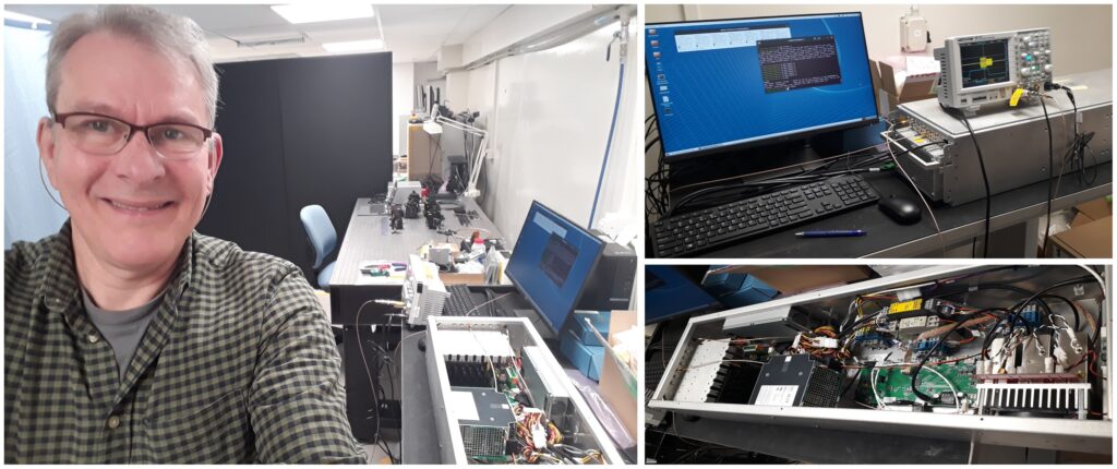
My part in Brighter has been to design/build the “AOM/AOD rack” which is the rack controlling the laser Modulation and Deflection using Acousto Optical elements. Also some adaptations to fit better into the Brighter setup. The AO technology is about 100 years old and consist of creating sound wave in a crystal to diffract light as the sound generates a kind of grating in the device. The frequencies involved are hundreds of MHz.
Could you tell us a little bit about the concrete work you’re involved in inside Brighter project?
Most of the AOM/AOD rack has direct counterparts in our pattern generators, so a small number of changes were required to adapt to Brighter. For example I made some scripts to ease the specific setup needed here. The optics, laser, alignment etc. are done by other people here.
What are the expected results?
The result is from our (Mycronic) part to deliver a system that can scan (or sweep) a laser beam while modulating it on and off, my parts are all the electronics and cables from the controlling computer up to the AOM/AOD devices.
What is the expected impact of the work you’re doing?
The SW we provide will take an bitmap image and produce the required actions from the AOM/AOD to create (by sweeping in time) lines – similar to an CRT. The vertical movement of the line is handled by GUF I believe.
How do you feel about being a part of this European Project?
It feels exciting, partly because it is so multidisciplinary and involves so many technologies, so I certainly risk to learn something new!
Anna Altshuler and Ruby Shalom-Feuerstein researchers from BRIGHTER project at Technion, Israel, publish a review article in the renowned journal Trends in Cell Biology, where they discuss open questions related to the adult stem cells, as their niche, dynamics, and pathology. The responses to these questions may have a crucial impact of the potential use of stem cells in regenerative medicine.
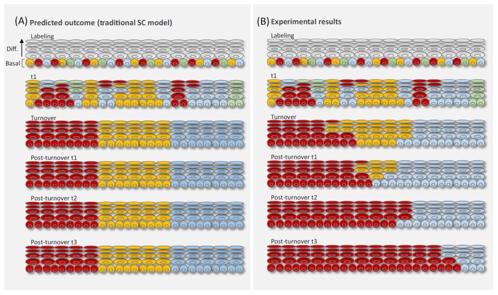
In the review article, entitled “Spotlighting adult stem cells: advances, pitfalls, and challenges”, authors focus on epithelial stem cells (SCs) and compare them to other SCs based on the discoveries made in the last years thanks to the advances in techniques like single-cell lineage tracing, live imaging, and genomics. They review the somatic SC paradigms of epithelial tissues, the significance of the new hypotheses, the similarities and key differences to other tissue SCs, and current challenges in the field.
Authors highlight that SCs are abundant in their niche, divide frequently, and that their differentiation is unpredictable. Beyond homeostatic regulation, another outstanding question concerns how the various SC types deal with tissue-specific challenges. They conclude the article stressing out that to see the benefits of SCs in the field of regenerative medicine it will be necessary to redirect research efforts towards human biology models to potentially allow reconstruction of functional tissues and genome editing of mutated SCs.
Francesco Pampaloni, coordinator of BRIGHTER Project at the Goethe-Universität Frankfurtam Main (GUF), recently published an editorial letter at the “International Journal of Molecular Sciences” talking about multi-modal and molecular imaging of cellular microenvironment and tissue development.
In this article, Pampaloni highlights the importance of properly imaging of individual cells inside their surrounding microenvironment for studies in fields like tissue engineering and bioprinting. Different technologies for the imaging of large three-dimensional cell cultures and even whole organs have been developed in recent years, and they are in constant evolution and improvement.
Different contrast modalities confer distinct advantages and drawbacks depending on the cell/tissue/organ to be observed, and the choice depends on the particularities of each case.
This article presents some of the most used techniques to image the cell microenvironment and the tissue with their pros and cons: the fluorescence microscopy and the label-free microscopy. However, the main subject explored in the article is the combination of both techniques in multi-modal systems. That can provide a deep insight into the cellular processes and interactions in highly heterogenous tissue microenvironments.
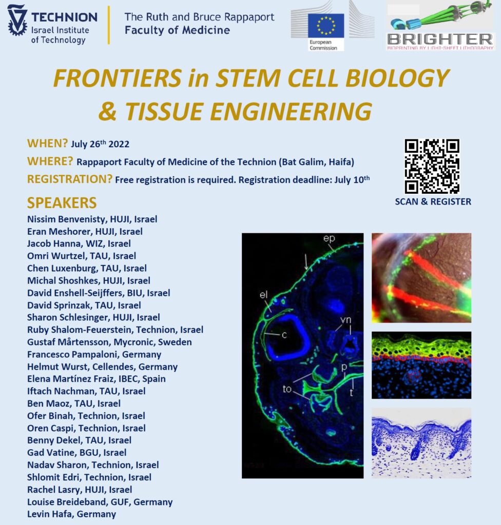
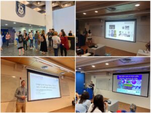
This week, the Faculty of Medicine of Technion, the Israel Institute of Technology welcomed the workshop “Frontiers in Stem Cell Biology & Tissue Engineering”, organized by the BRIGHTER project. Local and international renowned speakers covered the last advances in stem cells biology with emphasis on disease modelling and bioengineering.
During the 26th of July 2022, Haifa, in Israel, was the perfect place where several renowned researchers exchanged about Stem Cell Biology & Tissue Engineering. The BRIGHTER workshop “Frontiers in Stem Cell Biology & Tissue Engineering” could fortunately be held as an “in person” meeting after the pandemic, what greatly contributed to create an atmosphere of discussion and an ideal environment to share and acquire forefront knowledge in this topic.
Stem cells were first discovered in the 1950s-60s, and since researchers placed them at the top of the cellular differentiation pyramid, they were the object of uncountable research works that tried to understand their properties and regulatory mechanisms. The aim of this workshop was to contribute to this field summarizing the last advances in stem cells biology, with emphasis on subjects such as disease modelling and bioengineering. A wide range of topics were covered in the program, going from pluripotent and adult stem cells, the influence of the niche mechanobiology, cutting edge technologies of tissue modelling including advanced bioengineering and 3D bioprinting, individual cell imaging and genome-wide single cell analysis.
The event was organized by a BRIGHTER committee composed by one member of each consortium institution: E. Martínez (IBEC), G. Mårtensson (Mycronic), F. Pampaloni (GUF), H. Wurst (Cellendes) and R. Shalom Feuerstein (TECHNION). Moreover, some other BRIGHTER researchers were also there and could present their work. The workshop was divided in five sessions that grouped researchers talks in the following subjects: Pluripotency and reprograming; Development and adult stem cell niches; Advanced modelling with light sheet systems; Organoid and disease modelling; and Advanced modelling of morphogenesis and pathogenesis.
In this context, BRIGHTER researchers shared with the scientific community the core of the project: the development of a light sheet microscope based bioengineering technology that will allow high resolution modelling of complex stem cell niches and tissue structures with 3D tuneable matrix rigidities, while facilitating imaging individual stem cells in real time. Five talks by project members shaded light on this forefront topic: Ruby Shalom-Feuerstein: “Eye open on stem cells and their niche” Gustaf Mårtensson & Robert Eklund: “Shifting focus: moving existing technology between application spaces” Helmut Wurst: “Photocrosslinkable Biomimetic Hydrogels Based on the Thiol-ene Chemistry” Louise Breideband & Levin Hafa: “Imaging and bio-printing of 3D cell cultures with light sheet systems” Elena Martínez Fraiz: “Development of biomimetic models of tissue: guiding cellular self-organization through biofabrication techniques”
The workshop was a success and received more than 100 participants from different countries.
BRIGHTER project is funded by FET-open program from the European Commission under the grant agreement nº 828931.
Levin Hafa from GUF presented the advances of BRIGHTER Project at the second edition of the “EMBL-IBEC Conference on Engineering Multicellular Systems”, recently held in Barcelona.
Last 8th -10th of June Barcelona brought together renowned international researchers in fields focusing on how the engineering of multicellular living systems is driving our understanding of tissue and organ function. The “EMBL-IBEC Conference on Engineering Multicellular Systems” was centered in applications for disease modelling, drug screening, and tissue engineering.
Several talks and posters showed recent breakthroughs in stem cell biology, organ-on-chip assays, 3D bioprinting and cell mechanobiology, emphasizing in how strongly these technologies can influence our ability to design and assemble multicellular living systems, from organoids to embryos, and to impact in the medicine of the future.
In this context, Levin Hafa from GUF presented two posters about BRIGHTER project.
The first poster, signed by all partners of the project, summarized the objectives and results obtained in the project up to date. Levin presented the novel BRIGHTER’s bioprinting technique, which produces millimeter-sized objects in less than 3 minutes, using a digitally scanned light sheet. He also emphasized in the objective od producing human skin that will offer an alternative to animal testing for both pharmaceutical and cosmetics industries.
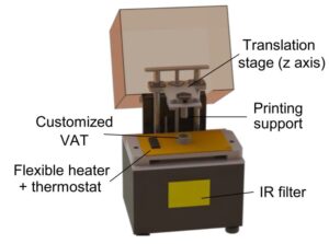
Additionally, he presented another poster about the democratization of the stereolithography 3D bioprinting: modelling the liver cancer microenvironment using a commercially available 3D printer, a work carried out at GUF by Louise Breideband. This work showed how researchers used a commercially available consumer stereolithographic 3D printer and modified it to a functioning 3D bioprinter. As a proof of concept, they were able to bio-print liver cholangiocarcinoma organoids.
Gustaf Mårtensson from MYCRONIC, explains in a video their role in the project as experts in optics, lasers and data handling. “By incorporating MYCRONIC’s lithographic semiconductor printing technology, we are able to manufacture cell-laden 3D constructs that resemble the complexity of human tissues”.
This video highlights the great opportunities of the 3D bioprinting technology to the biomedical field, concretely in the fabrication of human tissues that can be used for wound treating, drug testing and in the future, even for total organ replacement. Together with the other partners of BRIGHTER project, MYCRONIC develops a novel light-based patterning technology that can create this kind of tissues.
What MYCRONIC brings to BRIGHTER is a long experience in accurate acousto-optic patterning technology that can be combined with advanced cell laden bioinks that are light sensitive to be able to pattern very minor details down to the size of fan individual cell, in a quick and reliable way. MYCRONIC provides knowledge and manpower for system design, for pattern generators as well as for pattern generator optics, servo, data channel and system control software.
In a nutshell, they convert a three dimensional computer model of the biological tissue into a series of instructions for how to move a laser beam, and at the same time, control the light intensity of that laser beam, projected down to the size of a single human cell. One of the main advantages of this technology is that it’s highly scalable and cell friendly.
«I think it’s actually pretty amazing to take an existing technology that’s used in the world of electronics and transfer this to the realm of biology, where it has the opportunity to help so many people.» Gustaf Mårtensson
Brighter Project was present at the second Future 3D Additive Manufacturing, the 3DMM2O Conference in a poster and a flash talk by Angela Cirulli, PhD student at IBEC in Barcelona.
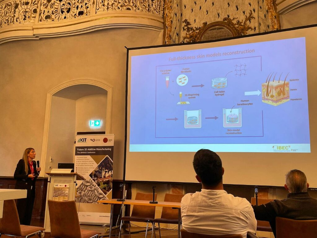
In the flash talk, Angela presented to an attentive and interested audience how BRIGHTER project aims to recreate skin-like tissue models resembling both the epidermal and the dermal compartments using photopolymerizable polymers. She focused her talk in the topic of full thickness skin models’ reconstruction and on light-based 3D Bioprinting systems. She highlighted the possibilities that offers the Digital Light Processing Stereolithography (DLP – SLA) to increase the scaffold dimensions needed both for mechanical characterization and to closely mimic the in vivo structures.
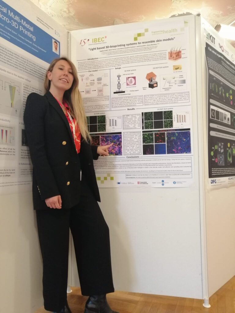
Besides the talk, Angela also presented a poster where she could go deeper and present more details of the project. This time she focused on the two light-based bioprinting approaches that have been applied to recreate skin-like tissue models. They first developed a skin model using a custom-made Direct Laser Writing (DLW) system working with visible light, and afterwards they scaled the size of the tissue-like constructs up to the cm scale by using a commercial Digital Light processing stereolithography (DLP-SLA) system. She also mentioned the next steps of BRIGHTER project towards the objective of developing a novel 3Dbioprinting technology to print organs and tissues at high resolution and speed using light.
A new scientific work led by Núria Torras and Elena Martínez, coordinator of the BRIGHTER project, shows a simple 3D bioprinting approach for the direct fabrication of advanced cell-laden tissue constructs by means of visible-light photopolymerization. The new approach allows the fabrication of cell-laden structures resembling the intestinal mucosa in a single printing step.
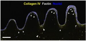
Confirmation of the 3D printed intestinal mucosa model by immunostainings of the main markers for both the epithelial and the stromal compartments showing cell distribution along the cross-sections of the 3D prints. Scale bar = 200 µm.
In recent years a lot of efforts have been made to produce gut engineered tissues representing reliable replicas of the in vivo tissue, including its particular architecture formed by finger-like protrusions called villi and invaginations called crypts. It is well known that the intestinal function relies not only on a healthy epithelium, but also on the stromal tissue (named lamina propria) that lays below. Therefore, to carry out consistent studies of inflammatory diseases, pathogen and microbiome interactions and even cancer, it is very important to have intestinal tissue models that resemble not only the epithelium but also the intestinal mucosa.
However, the intricate three-dimensional gut organization, together with complex biofabrication methods which entail a low cell survival rate and the high cost of some specialized equipment, limit the advances in the field of intestine tissue engineering. There is thus an urgent need to find easy fabrication methods of cell friendly complex structures, and in this scenario 3D bioprinting techniques can be a valuable tool to address this challenge, concretely light-based bioprinting techniques, due to their low cost, simplicity in use and versatility.
Custom 3D CAD model of our 3D bioprinter system.
Researchers from the Institute for Bioengineering of Catalonia (IBEC) led by Núria Torras and Elena Martínez, researchers from BRIGHTER Project, have developed a novel approach to produce fibroblast-laden crypt-villous structures by means of digital light processing (DLP) stereolithography (SLA). The work is available as a pre-print on bioRxiv repository. This technique photopolymerizes layer-by-layer bioinks, which can include a suspension of cells. By employing an optimized bioink formulation and suitable printing parameters, they were able to obtain a robust biofabrication approach that yields functional gut mucosa, with an excellent cell viability rate, accurate spatial resolution and high printing throughput.
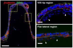
β-catenin (red) and ZO-1 (green) show epithelial cells polarization at different regions of the microstructures for both sample types (fibroblasts and Caco-2 cells). Sale bars = 100 µm, and 20 µm inserts.
Researchers developed a customized DLP-SLA 3D bioprinting system for the direct printing of tissue constructs using transparent, soft hydrogels, by means of visible-light photopolymerization. With this new approach, they were able to fabricate, in a single printing step, cell-laden structures resembling the intestinal mucosa and presenting the 3D architecture of the small intestine, including villi and crypts and the epithelial and stromal compartments.
“We have proposed a light-based 3D bioprinting approach as a feasible alternative for developing in vitro cell culture models recapitulating the native microenvironment of the in vivo tissue, thus contributing on providing alternatives beyond the current state-of-the-art”.
Núria Torras, first author of the study.
This new bioengineered tissue is compatible with conventional testing techniques, and the cost-effective DLP-stereolithography approach offers scalability, good resolution and fabrication speed, being a very good alternative to the existing in vitro systems.
The 11 of February was declared in 2015 as the International Day of Women and Girls in Science by the United Nations, and once again BRIGHTER researchers reinforced its commitment to this date participating with two talks and a hand-on activity oriented to young students.
In 2022 we celebrate the 7thInternational Day of Women and Girls in Science with the big challenge of eliminating gender stereotypes and long-standing biases that keep girls and women away from science. Worldwide, this day is dedicated to achieving full and equal access to science for women and girls. The different actions and events proposed aim to promote the empowerment of women and girls and their participation in science, further achieving gender equality.
This time, BRIGHTER project contributed with activities for young students from different age groups to help normalizing the presence of women in scientific fields.
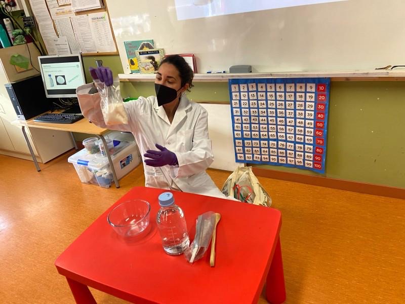
On one side, Nuria Torras, researcher at the Biomimetic systems for cell engineering group from Elena Martinez at IBEC in Barcelona, oriented her participation 30 6-7-years-old students from the school “El Vapor” in Terrassa, near Barcelona. First, she gave a talk titled “The scientists and their experiments” with an overview of what is a scientist, and talked about the research work she does in the laboratory in general and in the frame of BRIGHTER project.
Afterwards, she conducted a hands-on activity with the same students to explore the cell. During the activity “How I am? The cells and their functions” she explained basic concepts about the cell and the human organism.
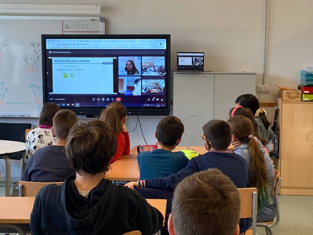
She gave an online talk to about 50 11-12 years-old students from the “La Canaleta” school at Vila-Seca, in the city of Tarragona. The talk was mainly centred in explaining her experience as a researcher and the path she has travelled to become a scientist. The audience was very interested and participated with interesting questions as:
How hard It was to become a scientist?
Has anyone tried to prevent you from being a scientist?
Why scientists change so many times amongst laboratories and countries?
Is it easy to have a family while being a scientist?
These questions reveal the importance of such kind of dissemination actions towards students, to help them see the scientific career as a “normal” career, and forget the stereotype of a crazy scientific man alone in his messy lab…
The European project BRIGHTER is developing a new technology to produce functional human tissues as an alternative to animal experimentation in the field of biomedical research.
This light-based 3D bioprinting technology fabricates tissues by patterning three-dimensional cell cultures. In the future, it could be even used to produce organs in the laboratory.
• The European project BRIGHTER is developing a new technology to produce functional human tissues as an alternative to animal experimentation in the field of biomedical research. • This light-based 3D bioprinting technology fabricates tissues by patterning three-dimensional cell cultures. In the future, it could be even used to produce organs in the laboratory.
Biomedical research progress made it possible to tackle diseases and make great medical advances in recent decades. Unfortunately, to the largest extent, experimental research required animal models to move forward. The European Union, through the European Association for Animal Research, strongly regulates animal research following the 3R principle. These are 1) Replacement: to avoid or replace the use of animals; 2) Reduction: minimise the number of animals used per experiment; and 3) Refinement: minimise animal suffering and improve welfare.
In line with European procedures, the BRIGHTER project (Bioprinting by light-sheet lithography: engineering of complex tissues with high resolution at high speed), coordinated by the Institute of Bioengineering of Catalonia (IBEC), was created precisely with the idea of contributing to reduce animal experimentation through the development of new solutions in 3D bioprinting. In this field, also known as tissue engineering or regenerative medicine, 3D printing techniques are increasingly used for biomedical purposes to produce bone, dental and cartilage prothesis. At BRIGHTER, researchers are fabricating human skin, a highly complex tissue, using light for 3D Bioprinting.
New BRIGHTER technology of 3D bioprinting
BRIGHTER is a European Union funded project coordinated by a group of experts from the Biomimetic systems laboratory for cell engineering from IBEC, led by Dr. Elena Martínez. Its main objective is developing and applying Light Sheet Bioprinting for the creation of complex and accurate in vitro models adequate for its use both in the pharmaceutical industry (testing of cosmetics and drugs) and in basic research, reducing animal experimentation.
To reach this end, researchers are developing a new 3D bioprinting technology based on patterned laser light sheet with which they intend to overcome some technical obstacles that currently limit the fabrication of complex human tissues.
«Our innovative 3D bioprinting system not only achieves tissues that are closer to the real ones, but it is also much faster than current systems, a fundamental factor to ensure the viability of the new tissues,» explains Professor Elena Martínez, coordinator of the European project.
Hydrogels, materials that form the base where the cells will grow and form the new tissue, are a key component in this technology. Hydrogels have properties resembling those found in the cellular environment in vivo (known as the extracellular matrix). This matrix surrounds the cells within the body, providing them with nutrients, tissue-like elasticity and stability. Another very relevant factor is that the entire process can be done in a personalized way, since patient cells can be used to construct the new tissue.
Laboratory printed skin
To fine-tune the new technology, BRIGHTER researchers are printing human skin, a tissue with a highly complex three-dimensional structure made up of multiple cell types and structures such as sweat glands and hair follicles.
On the one hand, the skin made with this new technology can be used as a substitute for animals both in the pharmaceutical and cosmetic industry, as well as in basic research laboratories, being a much more reliable system since it is made from human cells. On the other hand, it can help to respond to the demand for skin in medical interventions, for example, in burns or people suffering from different dermatological diseases.
The advantage of this new technology is that it allows to mould in detail the tissue being printed, which in the case of skin, is crucial since it is a dynamic tissue made up of several layers with different cell types and extracellular matrix composition. In addition, this technology makes it possible to generate vascularization of the tissue and include essential appendages such as the sebaceous (fat) and sweat glands, and the hair follicles that generate hair.
In order to «print» the skin, and for it to adopt its structure, shape and consistency, the researchers use advanced imaging techniques, which combine illumination with light sheets and high-resolution digital photomasks, that allow to pattern the cells inside the hydrogels. They do this by applying laser light directly onto a mixture of materials (hydrogels and cells), which also contains molecules that react to light. In this way, it is possible to mould the new tissue and create its 3D structure on demand, controlling the stiffness, shape and dimensions, thus generating three-dimensional tissues with complex geometry.
«We hope to be able to print a skin sample with an area of 1 cm2 and a thickness of 1 mm in approximately 10 min and with a cell viability of more than 95%, greatly improving current bioprinting conditions», concludes Dr. Núria Torras, postdoctoral researcher at IBEC.
Researchers from three top European research institutions (IBEC in Spain, Goethe Universität Frankfurt in Germany and the Technion center in Israel) participate in the BRIGHTER project, together with leading companies in the biotechnology sector (Mycronic in Sweden and Cellendes in Germany).
BRIGHTER project is funded by FET-open program from the European Commission under the grant agreement nº 828931.